

TNMU establishes Digi-CHange Lab digital laboratory
Ternopil National Medical University has established the “Digi-CHange Lab” digital laboratory as part of the Erasmus+ CBHE Digi-CHange project (“Digital Transformation and Curriculum Development for Healthcare Teams”).
This is an important step in the university’s efforts to advance the digital transformation of medical education and to expand opportunities for hands-on training for students and academic staff.
The “Digi-CHange Lab” is a new educational environment for integrating digital health tools into teaching and student training. In the lab, digital health technologies are used in the learning process to strengthen the practice-oriented approach to medical education.
Thanks to the new lab, students and faculty can master key areas of digital healthcare, including digital diagnostics, simulation-based learning, remote communication scenarios, AI-driven educational approaches, and the principles of responsible use of digital technologies in medicine.
The lab was established after the purchase and setup of equipment acquired with funding from the Digi-CHange project.
Digital technologies in medicine are opening up new possibilities for skin imaging, clinical case documentation, and improved training for future doctors. One of the modern tools for dermatological diagnostics is the “DermLite” dermatoscope, manufactured in the USA and purchased with Digi-CHange project funding. It is a compact optical device designed for detailed examination of skin lesions and skin structures.
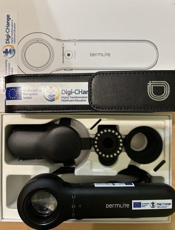
The “DermLite” dermatoscope can be connected to smartphones or tablets using appropriate adapters. This makes it possible to capture digital dermatoscopic images, store them for clinical documentation, use them in teaching, and compare results across repeated examinations.
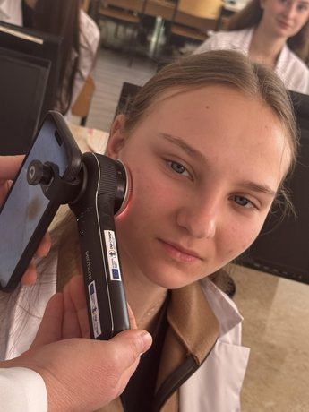
The dermatoscope’s optical system provides a magnified view of both surface and deeper skin structures, helping doctors assess the morphological features of pigmented lesions, vascular structures, and other dermatoscopic signs.
DermLite offers several lighting modes. Polarized light helps reduce glare and visualize structures beneath the skin’s surface, while non-polarized lighting allows for better assessment of surface features. This expands the possibilities of dermatoscopic examination.
Saved images can be used to compare the appearance of skin lesions across sequential examinations. This approach helps doctors track changes over time and determine the appropriate course of further evaluation.
The device’s portable design allows it to be used in a doctor’s office, during clinical examinations, and in practical classes. LED lighting illuminates the area under examination, eliminating the need for a separate lamp for each exam.
The use of “DermLite” in the educational process creates additional opportunities for the practical training of medical students. It can be used in classes on dermatology, visual diagnostics, and digital technologies in medicine, to demonstrate dermatoscopic features of various skin lesions, and to build skills in interpreting medical images.
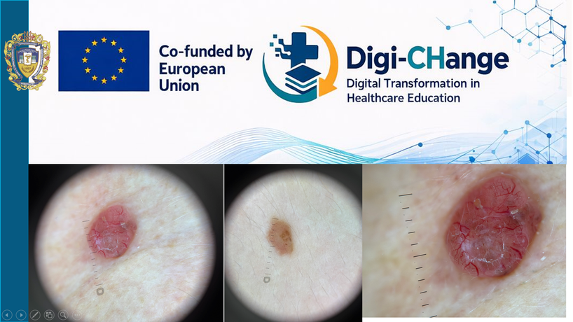
Digital images can also be useful for preparing teaching materials, discussing clinical cases, and conducting research. With proper compliance with personal data protection requirements, they can be used as material for developing and evaluating artificial intelligence algorithms, including machine learning methods for analyzing dermatoscopic images.
The “DermLite” dermatoscope is an example of combining optical technologies with digital tools that support the advancement of evidence-based medicine, improved diagnostic approaches, and the integration of innovation into medical education.
The use of modern dermatoscopic equipment helps bridge theoretical knowledge with practical skills and lays the groundwork for the further development of digital dermatological research at the intersection of medicine, information technology, and artificial intelligence.
Dermatoscopy is a supplementary method of clinical examination, as the results are assessed by a physician in light of the patient’s medical history and clinical presentation, with additional tests—such as a biopsy with histological examination—ordered as indicated.
It is in this context that the purchase of the “Grundium Ocus 40” digital microscope scanner, manufactured in Finland, with funding from the “Digi-CHange” project proved especially fitting.
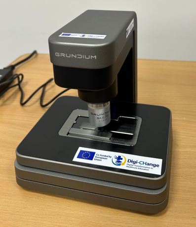
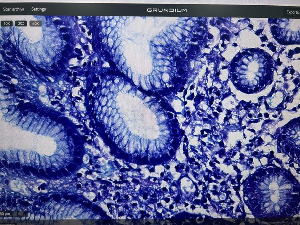
The digital microscope scanner is designed for high-quality digitization of histological and microscopic specimens, producing digital images that can be viewed, analyzed, stored, and used in educational and research activities.
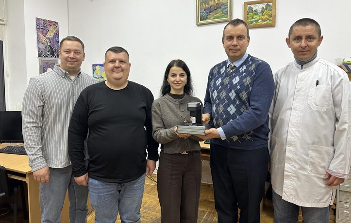
On October 19, 2026, training on the setup and effective use of the “Grundium Ocus 40” digital microscope scanner was conducted by Valerii Ostapets, an engineer at Global Biomarketing Group LLC (Kyiv).
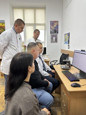
The “Grundium Ocus 40” microscope scanner makes it possible to create digital copies of microscope slides for detailed study of tissue structure, morphological research, and demonstrating microscope specimens to students and faculty. The use of digital images makes it easier for researchers to share materials, discuss results remotely, and build electronic teaching collections.
The scanner provides 40× magnification and a digital image resolution of 0.25 μm/pixel, allowing fine cellular and tissue structures to be examined. Automatic focusing and tissue detection simplify the scanning process.
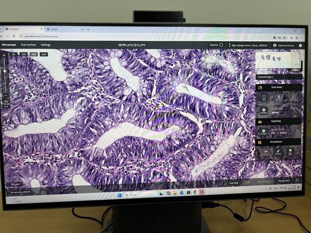
For the university, this equipment is especially important for advancing digital pathology, medical education, and AI-driven research. It can be used in the study of histology, cytology, and morphological tissue changes, in the analysis of biopsy materials, and in preparing digital image sets for developing and researching machine learning algorithms.
The “Digi-CHange Lab” digital laboratory expands the university’s capacity to integrate modern digital technologies into teaching and research, fostering interdisciplinary collaboration and the integration of medical knowledge with digital data analysis methods.
The “Digi-CHange Lab” will serve as a practical link between curriculum development and hands-on training in digital healthcare, supporting both the educational process and the professional development of staff, as well as the integration of digital health topics into the curricula of I. Horbachevsky Ternopil National Medical University.
By establishing the “Digi-CHange Lab” digital laboratory, the university affirms its high standards in preparing future healthcare professionals to work in modern digital medical laboratories, where information technology serves as a supporting tool but does not replace physicians’ professional judgment, ethical responsibility, teamwork, and patient-centered care.
Information and photos prepared by Digi-CHange project coordinator Professor Andrii Sverstiuk and Solomiia Kramar, associate professor at the Department of Histology and Embryology.